|
I am a Ph.D. student at Stanford University where I work on computational magnetic resonance imaging and artificial intelligence. I am advised by Shreyas Vasanawala and funded by the NSF Graduate Research Fellowship. I did my undergrad at University of Southern California where I worked with Krishna Nayak on cardiac MRI for a few years. I've also interned at the National Institutes of Health and GE Healthcare. This summer I will be working at Apple AI Research |
|
|
|
|
Broadly, I'm interested in developing signal processing and artificial intelligence approaches to solve problems in computational sensing. Currently, I focus on upstream AI methods to improve pediatric magnetic resonance imaging with respect to speed and diagnostic accuracy. Representative papers are highlighted. |
|
Christopher Sandino, Peng Lai, Shreyas Vasanawala, Joseph Cheng Presented at ISMRM 2019 (Magna Cum Laude). Submitted to MRM, 2019. Designed a convolutional neural network architecture to reconstruct dynamic MR images from highly undersampled raw data by jointly leveraging physics-based signal models and deep 3D priors. Using this technique, standard MRI scans which once took 5-6 breath-holds now take a single breath-hold allowing for faster and higher accuracy assessment of heart function. |
|
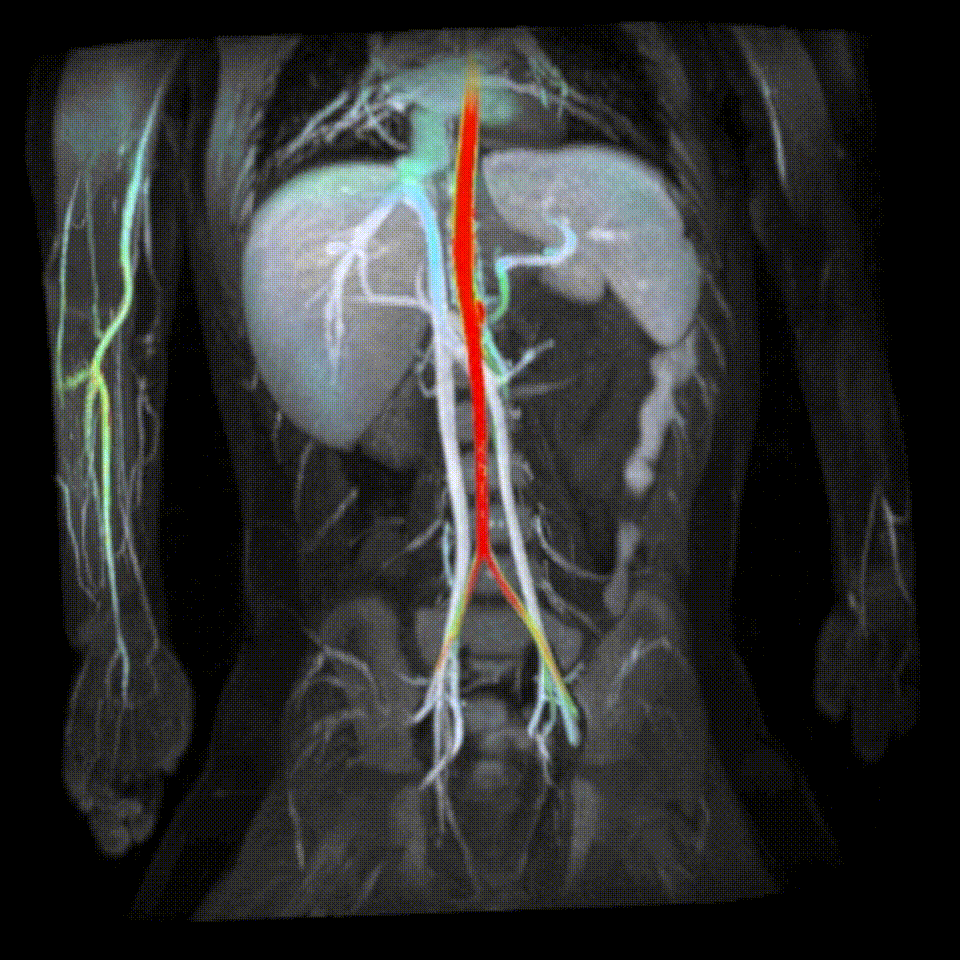
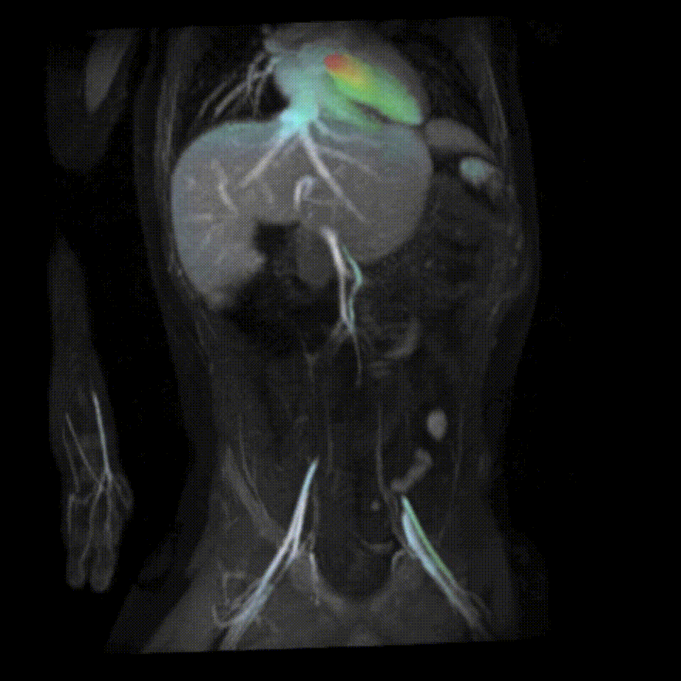
Christopher Sandino, Frank Ong, Joseph Cheng, Marc Alley, Shreyas Vasanawala Presented at ISMRM 2018 (Magna Cum Laude). Developed a motion-robust MRI acquisition scheme to enable measurement of velocity vector fields as a function of 3D space and time. We leverage low-rank compressed representations and a stochastic algorithm to reconstruct 100GB datasets acquired from each scan. Our technique has enabled fast, free-breathing, and comprehensive abdominal MRI exams for pediatric patients in as little as 5 minutes. |
|
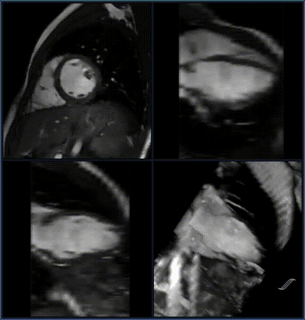
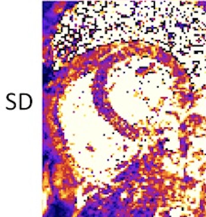
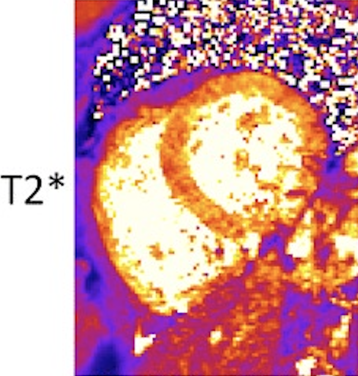
Christopher Sandino, Peter Kellman, Andrew Arai, Michael Hansen, Hui Xue Presented at ISMRM 2015. Published in JCMR, 2015. Developed a more robust method for estimating T2*, a magnetic tissue parameter which can be used to identify iron overload in the heart. We also formulated a method to derive "confidence" maps using noise statistics measured with a short and simple calibration scan. This technique is implemented in medical image reconstruction framework, Gadgetron, and is being used at over 30 clinical sites worldwide. |
|
Template borrowed from Jon Barron. |